 University Journal of Surgery and Surgical Specialities
ABDUCENT NERVE PALSY- A RARE CASE REPORT
Abstract
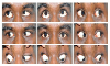
A 34-year old male who developed binocular horizontal double vision in right gaze was found to have an isolated sixth nerve palsy in right eye secondary to sub-acute bleed involving Pons due to solitary small Cavernous malformation. The natural history of Cavernous malformation and the mechanism by which hemorrhage of these vascular lesions may produce minimal neurological signs, including isolated ocular motor cranial nerve palsies, is discussed. Magnetic resonance imaging (MRI) that includes susceptibility-weighted sequences leads to their accurate diagnosis.
Full Text:
PDFReferences
Batra S, Lin D, Recinos PF, Zhang J, Rigamonti D. Cavernous malformations: natural history, diagnosis, and treatment. NatRev Neurol. 2009;5:659–670.
Curling OD Jr, Kelly DL Jr, Elster AD, Craven T. An analysis of the natural history of cavernous angiomas. J Neurosurg.1991;75:702–708.
Fritschi J, Reulen J, Spetzler R, Zabramski J. Cavernous malformations of the brain stem. A review of 139 cases. ActaNeurochir (Wien). 1994;130:35–46.
Robinson JR, Awad IA, Little JR. Natural History of the cavernous angioma. J Neurosurg. 1991; 75:709-714.
Flemming K, Link M, Christianson T, Brown R. Prospective hemorrhage risk of intracerebral cavernous malformations. Neurology. 2012;78:632-636.
Key words:
Cavernous malformations, Cavernous angioma, Abducent nerve palsy.
Refbacks
- There are currently no refbacks.

This work is licensed under a Creative Commons Attribution-NoDerivatives 4.0 International License.
An Initiative of The Tamil Nadu Dr MGR Medical University