 University Journal of Surgery and Surgical Specialities
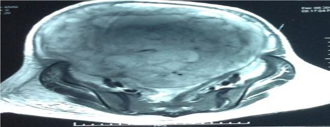
A Diagnostic dilemma for the gynaecologist- an ovarian mass with an unexpected twist- a rare case of leiomyosarcoma arising from retroperitoneum
Abstract
Full Text:
PDFReferences
Mettlin C, Priore R, Rao U. results of the national soft tissue sarcoma registry. J Surg Oncol. 1982;19:224 – 7 (PubMed)
Issac R Francis, Richard H Cohan, Datla G K Varma and Vernon K Sondak. Cancer Imaging 2005;5(1):89-94
Retroperitoneal tumours in the pelvis : a diagnostic challenge in gynecology . Wei-Wei Wee-Stekly and Micheal David Mueller . front . surg ., 5 Dec,2014
Gatita CE, Georgescu I, Nemes R. difficulties in diagnosis of primitive retroperitoneal tumours. Curr Health Sci J(2010) 36(3):132-5 (PubMed)
Huemen MT, Herman JM, Ahuja M. management of retroperitoneal sarcomas.Surg Clin North Am (2008) 88:583-97. Doi:10.1016/j.suc.2008.03.002
Osman S, Lehnert BE, Elojeimy S, Cruite I, Manneli L, Bhargava P, et al. A Comprehensive review of retroperitoneal anatomy, neoplasms and pattern of disease spread. Curr Probl Diagn Radiol (2013) 42(5): 191-208. Doi:10.1067/j.cpradiol.2013.02.001
Elsayes KM, Staveteig PT, Narra VR, Chen ZM, Moustafa YL, Brown J. Retroperitoneal masses: magnetic resonance imaging findings with pathologic correlation. Curr Probl Diagn Radiol (2007) 36: 97-106. Doi:10.1067/j.cpradiol.2006.12.003
Shanbhogue AK, Fasih N, Macdonald DB, Sheikh AM, Menias CO, Prasad SR. Uncommon primary pelvic retroperitoneal masses in adults: a pattern based imaging approach. Radiographics (2012) 32:795-817.doi:10.1148/rg.323115020. (Pubmed)
Center for Disease Control Manual. Guide: Safety management, no.CDC-22, Atlanta, GA.
Pregnant Women Presenting with a Gross Retroperitoneal Mass: Surgical Treatment with Caval Replacement. Roberto bertini, Nazareno Suardi, et al. European association of urology, European Urology 54 (2008)677-680.
Leiomyosarcoma of the rectum mimicking Primary Ovarian Carcinoma: a case report Yung-Taek Ouh, Jin Hwa Hong, Kyung-Jin Min, Kyeong-A So, Jae Kwan Lee. Journal of Ovarian Research 2013 6:27 doi:10.1186/1757-2215-6-27
A Case of Retroperitoneal Liposarcoma after delivery with expression of estrogen receptor: a case report Hiroaki Kakashima, Yoshio Yamasaki, Yasuyuki Yoshida, et al. Int j Surg Case Rep.2015;7:99- 103.
Li X.-Q., Hisaoka M., Hashimoto H. Expression of estrogen receptors alpha and beta in soft tissue sarcomas: immunohistochemical and molecular analysis. Pathol. Int. 2003;53:671-679.( Pubmed)
Refbacks
- There are currently no refbacks.

This work is licensed under a Creative Commons Attribution-NoDerivatives 4.0 International License.
An Initiative of The Tamil Nadu Dr MGR Medical University