 University Journal of Surgery and Surgical Specialities
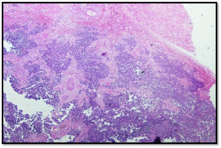
A rare presentation of ovarian tumour
Abstract
Primary neuroectodermal tumours of ovary are rare monophasic teratomas composed exclusively of neuroectodermal tissue. Most tumours were diagnosed in third and fourth decade of life1. Microscopically they are identical to neuroectodermal tumours of the CNS. They had usual presenting symptom of abdominal pain or abdominal lump, the tumour which varied from cystic to solid in consistency and size ranged from 4 – 20 cm in diameter2. Our case a 50 yr old post menopausal lady presented with abdominal pain was evaluated, Ultrasonogram or MRI, revealed a huge cystic ovarian mass without ascites3. Ovarian tumour markers were all within normal range. Underwent Total abdominal hysterectomy, bilateral salphingo-oopherectomy, and omentectomy. Histopathological examination showed neuroectodermal tumour of ovary.
Full Text:
PDFReferences
Histol Histopathol.2008Jun;23(6):765-71.doi:10.14670/HH
-23.765.Neuroectodermal ovarian tumors: a brief
overview. Morovic A1, Damjanov I.
1993 Aug;17(8):764-78.Primary neuroectodermal tumors
of the ovary. A report of 25 cases.Kleinman GM1, Young
RH, Scully RE.
Saudi Med J. 2008 Mar;29(3):444-6.Primitive
neuroectodermal tumor of the ovary.Anfinan NM1, Sait KH,
Al-Maghrabi JA.
The Korean Journal of Pathology 1999;33(8): 631-635.
Primitive Neuroectodermal Tumor of the Ovary: A case
report.Chan Kwon Jung, Eun Sun Jung, Youn Soo Lee,
Byung Kee Kim, Sun Moo Kim
Kawauchi, Shigeto M.D.; Fukuda, Toshiro M.D.;
Miyamoto, Shingo M.D.; Yoshioka, Jun-Ichi; Shirahama,
Syuya; Saito,Toshiaki M.D.; Tsukamoto, Naoki M.D.
peripheral primitive neuroectodermal tumour of the
ovary confirmed by CD99 immunostaining.
Refbacks
- There are currently no refbacks.

This work is licensed under a Creative Commons Attribution-NoDerivatives 4.0 International License.
An Initiative of The Tamil Nadu Dr MGR Medical University