 University Journal of Surgery and Surgical Specialities
A Rare Case of Hypertropia: A Case Report
Abstract
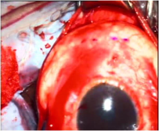
A 33 year old female presented to us with squinting of left eye since birth. On examination patient had large angle hypertropia with exotropia in left eye which was a diagnostic challenge. Imaging showed reduction in volume of inferior rectus muscle in left eye. Intraoperatively inferior rectus muscle was found to be absent. The surgical correction resulted in excellent alignment in primary position. Hence high index of suspicion is required to consider inferior rectus aplasia. This case is presented for its rarity and surgical expertise to achieve the best possible outcome.
Full Text:
PDFReferences
Pei-Yu Lin and May-Yung Yen. “Congenital absence of bilateral inferior rectus muscles: a case report”. Journal of pediatric ophthalmology and strabismus 34.6 (1997): 382- 384.
SB Ozkan., et al. “Hypoplastic inferior rectus muscle in association with accessory extraocular muscle and globe retraction”. Journal of AAPOS 11.5 (2007): 488-490.
Diamond GR, Katowitz JA, Whitaker LA. Variations in extraocular muscle number and structure in craniofacial dysostosis. Am J Ophthalmol 1980;90:416-8.
Bhate M, Martin FJ: Unilateral inferior rectus hypoplasia
in a child with Axenfeld-Rieger syndrome. J AAPOS 2012;16:304–306.
Taylor RH, Kraft SP. Aplasia of the inferior rectus muscle.
A case report and review of the literature. Ophthalmology 1997;104:415-8.
Astle WF, Hill VE, Ells AL, Chi NT, Martinovic E.
Congenital absence of the inferior rectus muscle—diagnosis and management. J AAPOS 2003;7:339-44.
Refbacks
- There are currently no refbacks.

This work is licensed under a Creative Commons Attribution-NoDerivatives 4.0 International License.
An Initiative of The Tamil Nadu Dr MGR Medical University