 University Journal of Surgery and Surgical Specialities
A Retrospective Study on variations In Chin-Throat Angle in Different Skeletal Malocclusions
Abstract
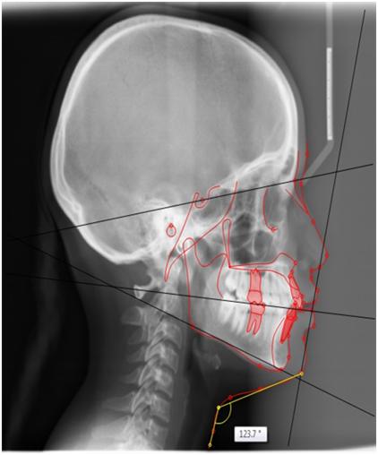
OBJECTIVE: To assess the variations in Chin Throat Angle in different skeletal malocclusions in local population reported to Madha dental college and hospital.MATERIALS & METHODS: Profile photographs and lateral cephalograms of 102 adults aged between 18-28 years were taken, out of which 75 were class I, 23 were class II and 4 were class III malocclusions. All the samples were digitalized and standardized and ANB and Chin Throat Angles were measured. The pre-treatment measurements were compared in between these three groups. The Statistics included normality test, Kolmogorov-Smirnov and Shapiro-Walks tests, parametric test, one way ANOVA and independent t-test for group differences.RESULTS: Chin Throat Angle was increased in class II group than that of class I and III groups. Significant differences (P< 0.05) were observed for ANB and Chin Throat Angle between Classes I/II/III as (<0.01) & (<0.036) respectively on lateral cephalograms. On profile photographs Chin Throat Angle between the three classes were significant (<0.027).CONCLUSION: The Chin Throat Angle analysis is essential for the orthodontist’s awareness of treatment planning and can also be used for explaining to the patient. Thus not only frontal photographs but also lateral cephalograms along with profile photographs plays an important role in diagnostic purposes.
Full Text:
PDFReferences
Jacobson A, Vlachos C. Soft tissue evaluation. In: Jacobson A, Jacobson RL, eds. Radiographic Cephalometry—From Basics to 3-D Imaging, 2nd edition. Chicago, Ill: Quintessence Publishing Co; 2006:205–217.
Subtelny J. A longitudinal study of soft tissue facial structures and their profile characteristics, defined in relation to underlying skeletal structures. Am J Orthod. 1959;45:481–507.
Burstone CJ. Integumental contour and extension patterns. Angle Orthod. 1959;29:93–104.
Ricketts RM. Esthetics, environment, and the law of lip relation. Am J Orthod. 1968;54:272–289.
Mommaerts MY, Marxer H. A cephalometric analysis of the long-term, soft tissue profile changes which accompany the advancement of the mandible by sagittal split ramus osteotomies. J Cranio-Maxillofac Surg. 1987;15:127–131.
Bergman RT. Cephalometric soft tissue facial analysis. Am J Orthod Dentofacial Orthop. 1999;116:373–389.
Ellenbogen R, Karlin JV. Visual criteria for success in restoring the youthful neck. Plast Reconstr Surg. 1980;66:826–837.
Moreno A, Bell WH, You ZH. Esthetic contour analysis of the submental cervical region: a study based on ideal subjects and surgical patients. J Oral Maxillofac Surg. 1994;52:704– 713; discussion 713–714.
Sommerville JM, Sperry TP, BeGole EA. Morphology of the
submental and neck region. Int J Adult Orthod Orthognath
Surg. 1988;3:97–106.
Naini FB, Cobourne MT, McDonald F, Wertheim D.
Submental-cervical angle: perceived attractiveness and
threshold values of desire for surgery. J Oral Maxillofac
Surg. 2016;15:469–477.
Marino H, Galeano EJ, Gondolfo EA: Plastic correction of double chin; importance of the position of the hyoid bone. Plast Reconstr Surg 31:45. 1960.
Worms F. lsaacson R. Speidel T: Surgical orthodontic treatment planning-Profile analysis and mandibular surgery. Angle Orthod 46:1, 1976.
Ellenbogen R, Karlin J: Visual criteria for success in restoring the vouthful neck. Plast Reconstr Sum 66:826. 1980.
Sommerville JM. Sperry TP. BeGole EA: Morphology of the
submental and neck region, Int J Adult Orthod Orthognath
Sure 3~97. 1988.
Mitz V: Peyronie M: The superficial mu & oaponeurotic system (SMAS) in the parotid and cheek. Plast Reconstr Surg 58:80. 1976.
Lindner HH: The anatomy of the fasciae of the face and neck with particular reference to the spread and treatment of intraoral infection (Ludwig’s) that have progressed into adjacent facial spaces. Ann Surg 204:705. 1986.
Vistnes LM. Souther SC: The platysma muscle. Anatomic considerations for esthetic surgery of the anterior neck. Clin Plast Surg 10:441. 1983.
Cardoso de Castro C: The anatomy of the platysma muscle. Plast Reconstr Surg 66:680. 1983.
Standardized portrait photography for dental patients
Lewis Claman, DDS, MS, Daniel Patton, BA, RBP, and Robert Rashid, DDS Columbus, Ohio, AM J ORTHOD DENTOFAC ORTHOP 1990;98:197-205.)
Chin-throat anatomy: Normal relations and changes following orthognathic surgery and growth modification Ramzi V. Haddad; Joseph G. Ghafari. (Angle Orthod.0000;00:000–000)
Refbacks
- There are currently no refbacks.

This work is licensed under a Creative Commons Attribution-NoDerivatives 4.0 International License.
An Initiative of The Tamil Nadu Dr MGR Medical University